COMPOUND MICROSCOPE::
microscope·@abdulhanankhan·
0.000 HBDCOMPOUND MICROSCOPE::
-1.jpg)
-1.jpg)
-1.jpg)
-1.jpg)
-1.jpg)
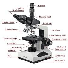
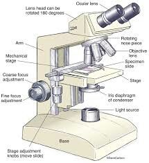
COMPOUND MICROSCOPE::
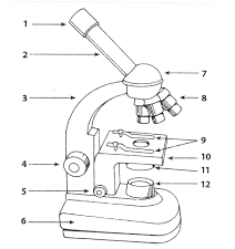
Whenever high magnification is desired, a pompound microscope is used. It consists of two convex lenses, an object lens of very short focal length and an eye-piece of comparatively longer focal length.
The ray diagram of a compound microscope is given blew.
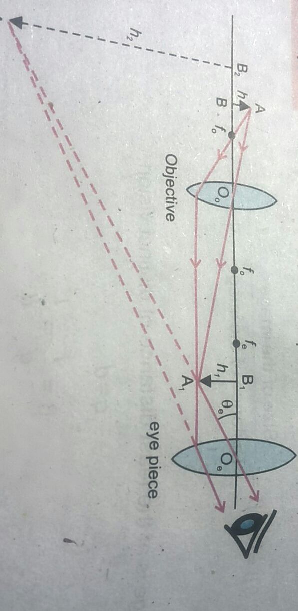
The object of height "h" is placed just beyond the principal focus of the objective.This produces a real, magnified image of hight "h1" of the object at a place situated within the focal point of the eye-piece.........
It is ustomary to refer the values of "M" as multiples of 5, 10, 40 etc., and are marked as x5, x10, x40 etc., on the instrument.
The limit to which a microacope can bevused to resolve details, depends on the width of the objective.A wider objective and use of blue light of short wavelength produces less diffraction and allowa more details to be seen.
If you are Like my artical
Follow me
And
Upvote me
Thanks for read my artical.